Surgical Guide - For Precision and Perfection
Digital Impressions
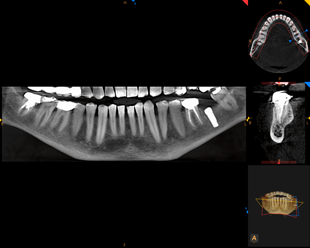
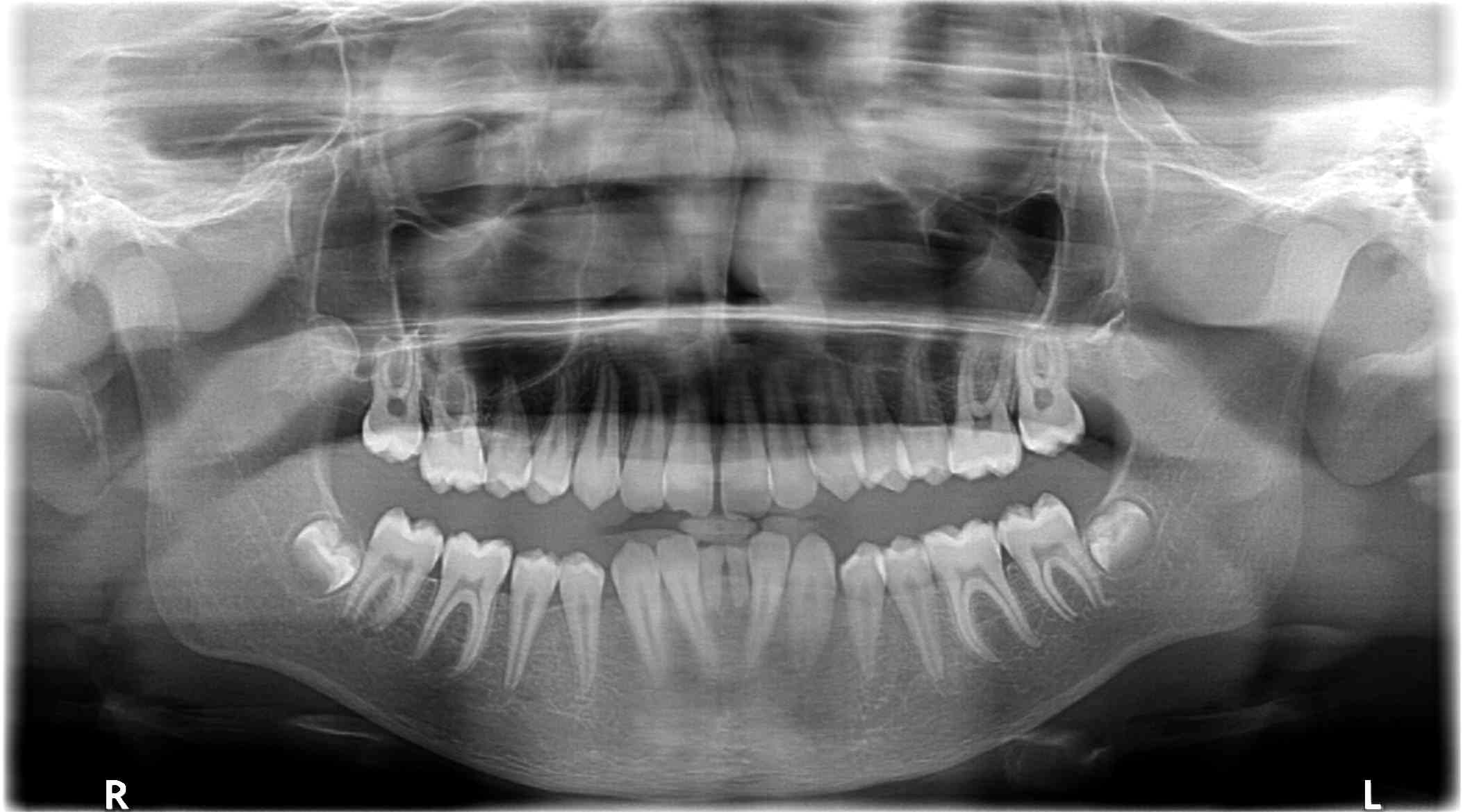
OPG
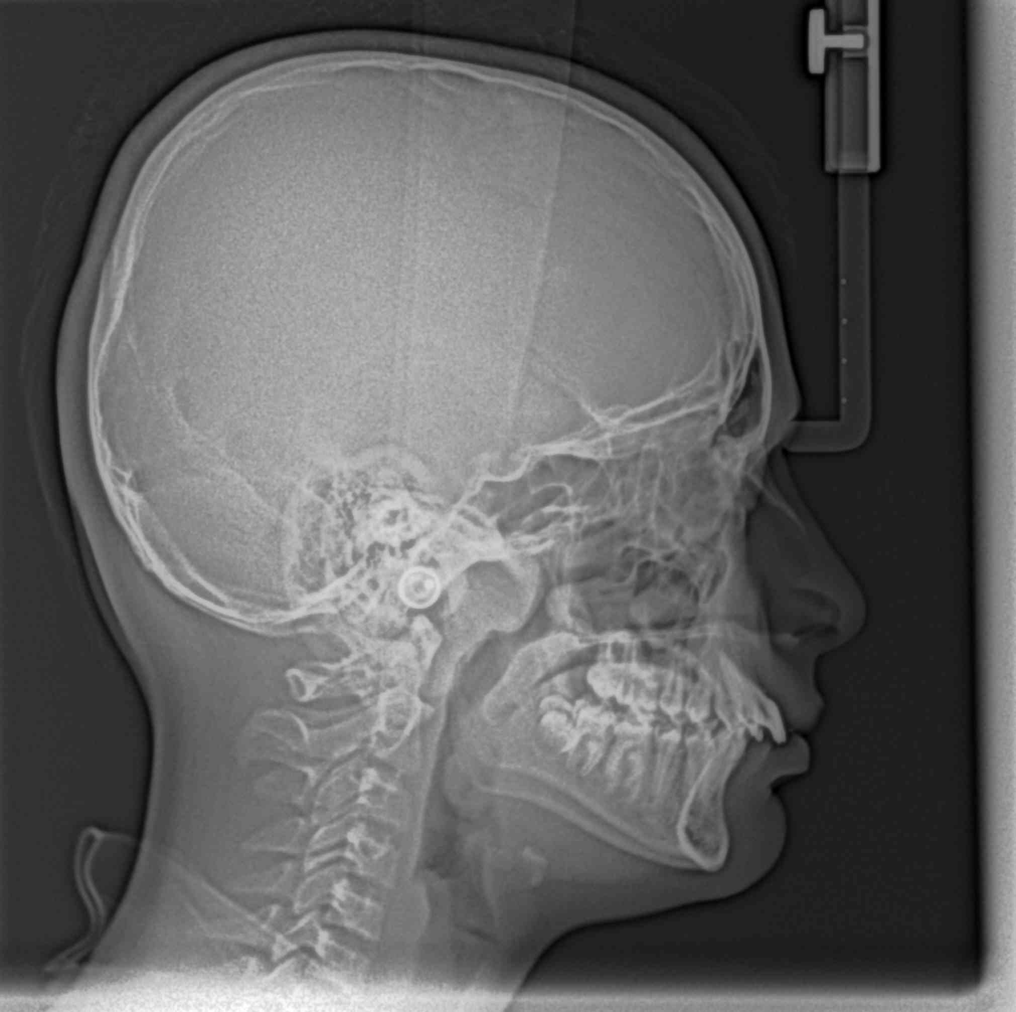
LATERAL
CEPHALOGRAM
ANTEROPOSTERIOR VIEW
HAND WRIST RADIOGRAPH
OCCLUSAL VIEW
TMJ[OPEN AND CLOSE]
PNS
SMALL FIELD OF VIEW ( FIELD OF 5X5 cms)
MID FIELD OF VIEW ( FIELD OF 8X4 OR 8X8 cms)IT IS EITHER FULL MAXILLA/MANDIBLE/BOTH IN A SINGLE SCAN
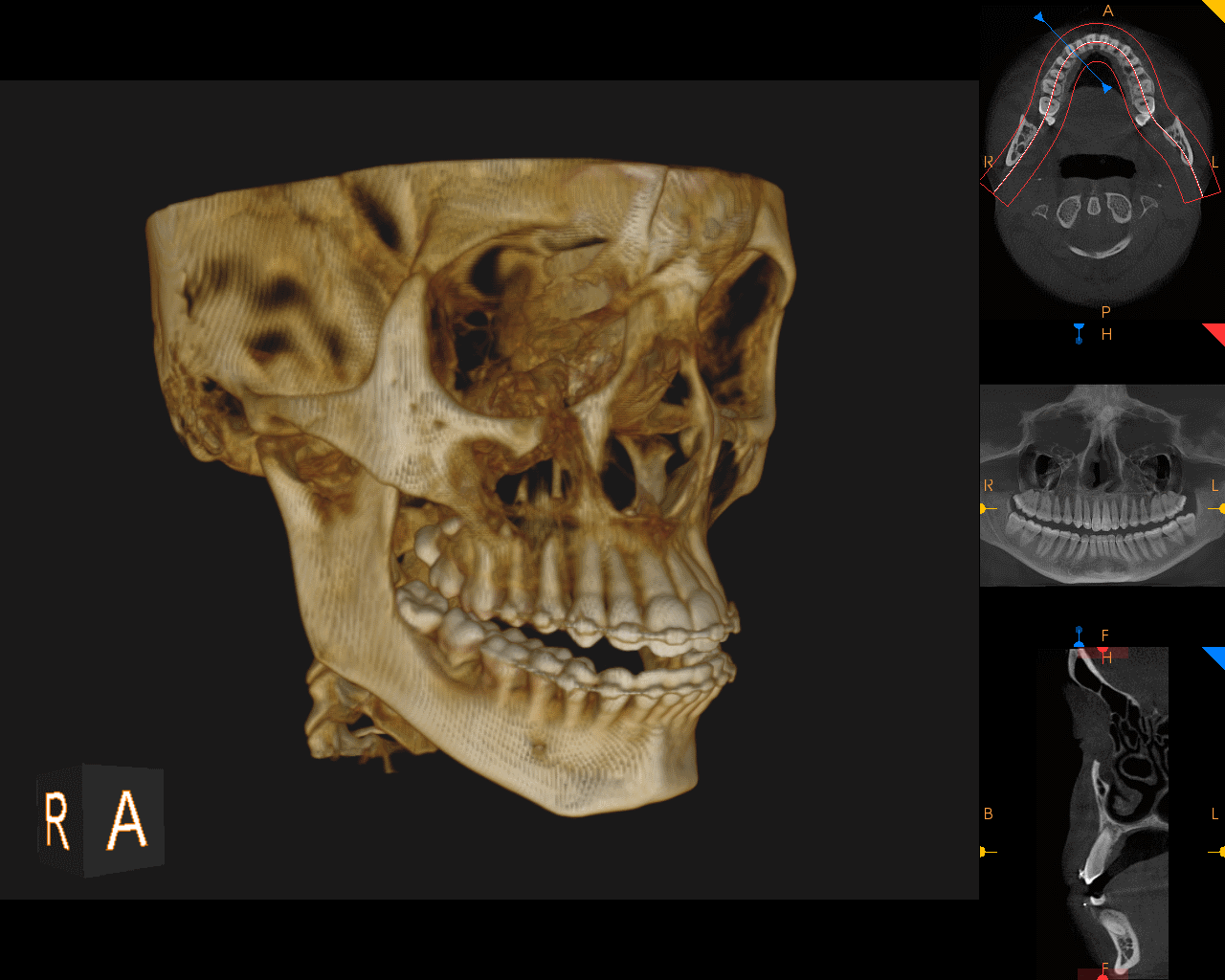
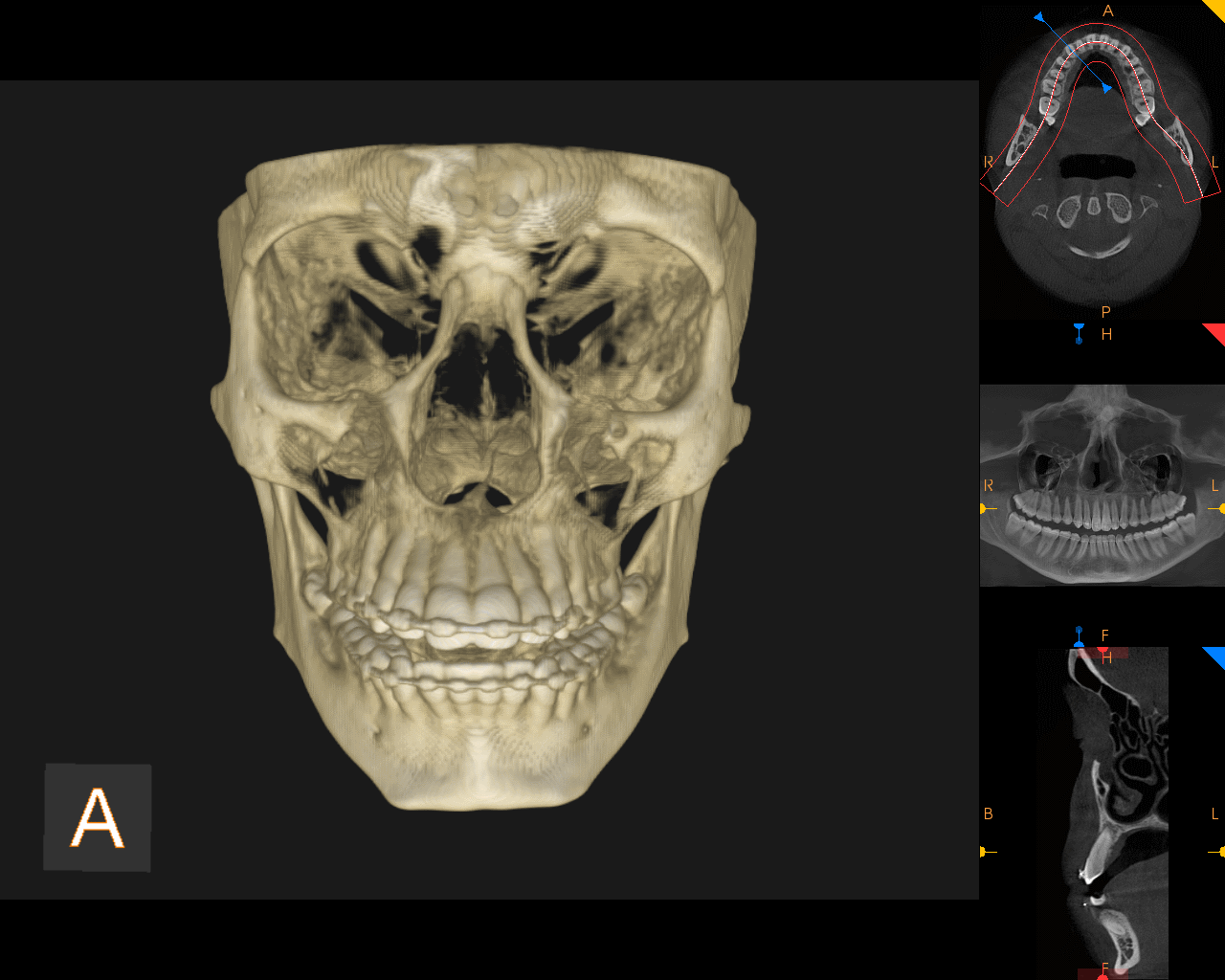
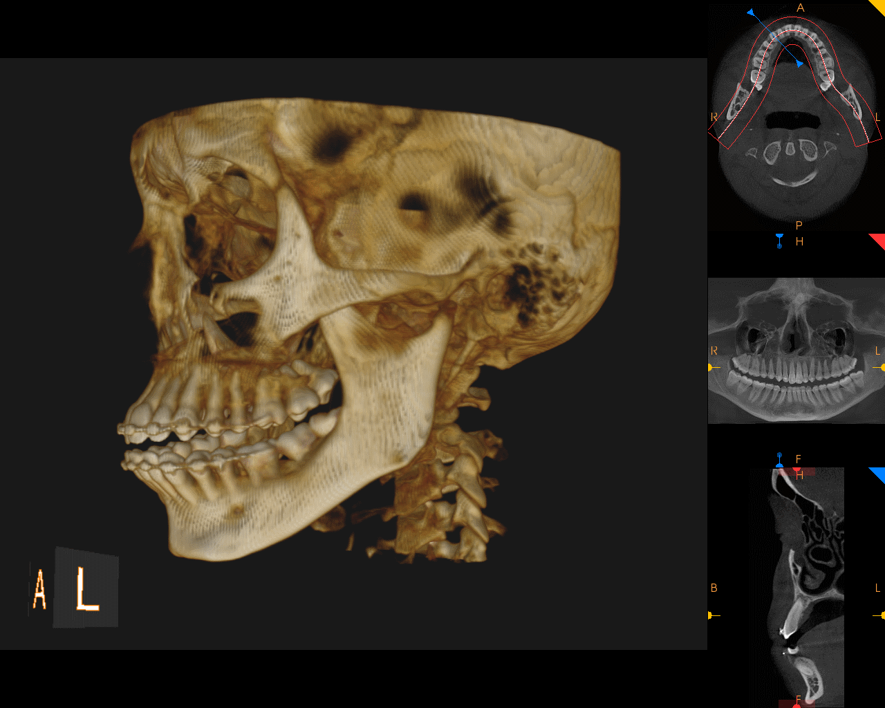
LARGE FIELD OF VIEW (FIELD OF 14X17 cms)SCAN COVERING FULL SKULL
CBCT OF TMJ
CBCT OF SINUSES
RESEARCH WORK INVOLVING CBCT SCANS
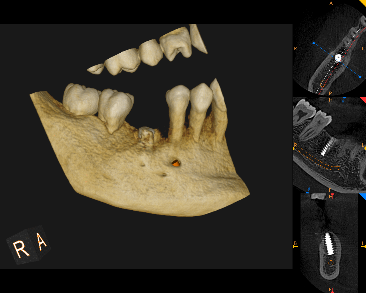
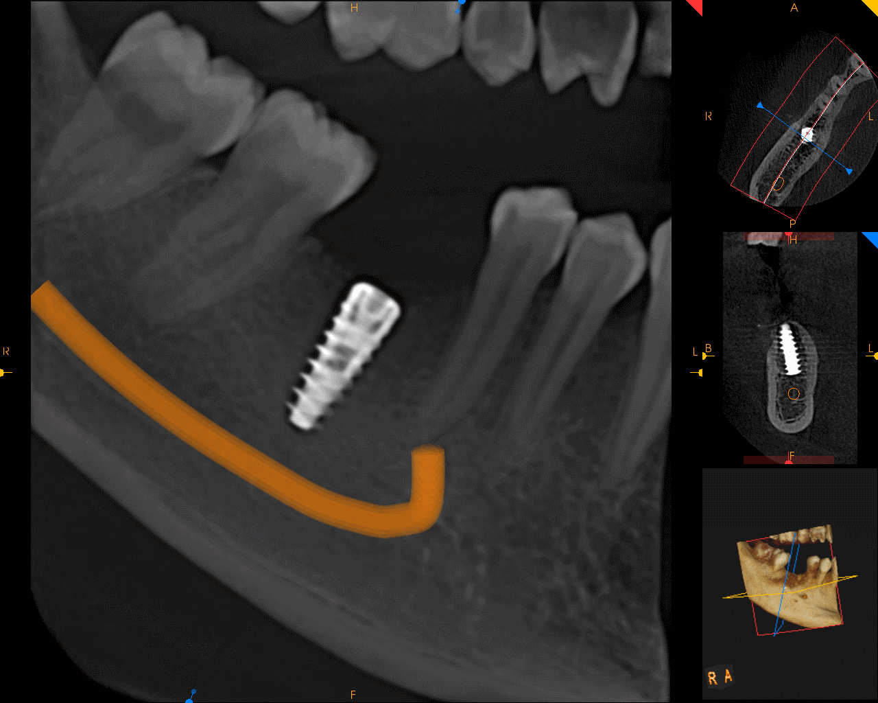
SCANS FOR SURGICAL GUIDE FABRICATIONS
RIGHT AND LEFT TMJ
RIGHT AND LEFT OPEN AND CLOSED
MOUTH TMJ
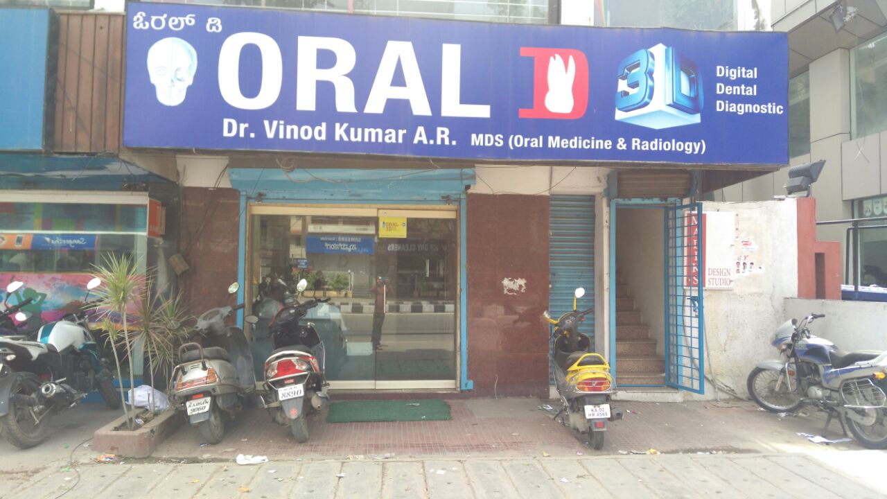
Established in the year 2010. The first Radiology practice to introduce Cone Beam CT (CBCT) technology to Bangalore….
The emerging "standard of care" for the diagnostic assessment of the bony components of the face.
The centre is managed by efficient oral and maxillofacial radiologists.
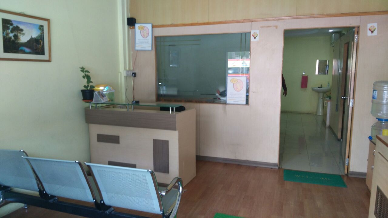
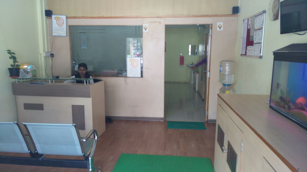
Highly hospitable environment for patients,high quality images and detailed radiologic reports will help referring dentist with improved treatment planning and more efficient patient care
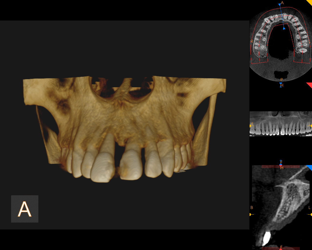
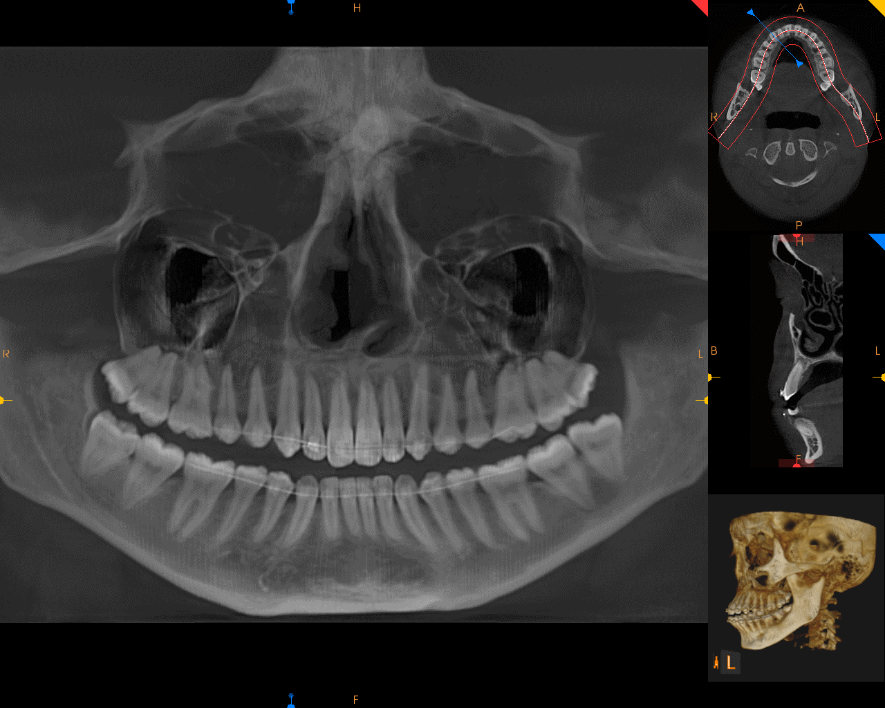
2D and 3D imaging facilities available
Full 3D volumetric scan — multiple images are generated in various planes enabling multi-dimensional assessment, accurate diagnoses, improved treatment planning and more efficient patient care.
• IMPLANT assessment
Detailed anatomy of the area in 3 dimensions Length and width of the available bone in the desired area Virtual implant planning
• SURGERY
In evaluating 3rd molar, canine and other impactions Trauma assessments Visualizing pathological lesions
• ORTHODONTICS
Position of impacted canine Assessing the reasons for Delayed movements of the teeth
• ENDODONTICS
Anatomy of root canals Extra root canals Calcifications Vertical and horizontal fractures Failed root canal treatment assessments
• TMJ
In diagnosing any bony pathology
Different Scanning Images available

Different equipment/facilities available